
Are We Underestimating Dynamic Compression in Cervical Myelopathy?
An interesting second-opinion case in clinic today highlighted the potential value of flexion-extension MRI.
A patient underwent C5–6 ACDF approximately eight years ago for severe myelopathy and did very well. He now has progressively worsening myelopathic symptoms. His neutral MRI showed only moderate stenosis, and another surgical team had recommended against further surgery.
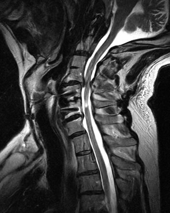
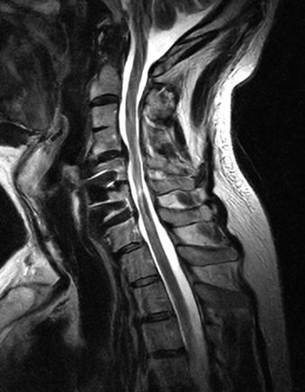
I initially agreed—the static imaging did not seem to explain his deterioration. But his symptoms were worsening and examination was clearly myelopathic.
Flexion-extension MRI demonstrated significantly greater dynamic cord compression than was apparent on the neutral study.
It raises a few questions for spine surgeons:
- Do you routinely use flexion-extension MRI when the clinical examination and static MRI don’t correlate?
- How do you incorporate dynamic compression into surgical decision-making?
- This patient also has a congenital C3–4 fusion and prior C5–6 ACDF, leaving C4–5 as a mobile segment between two fused levels. How would you approach the biomechanics and surgical strategy in this situation?
Cases like this reinforce that cervical myelopathy is ultimately a clinical diagnosis, while a neutral MRI captures only one position of a dynamic cervical spine.
Would love to hear how others are using dynamic imaging in their practice and whether it has changed management.